Scoliosis assessment has traditionally relied on radiographic imaging to quantify spinal deformity. While radiographs remain essential for diagnosing and monitoring curve progression, they provide a static view of a condition that strongly affects dynamic function.
Scoliosis is not only a structural spinal deformity but also a functional disorder that alters posture, balance, and gait mechanics.
These insights are supported by the study “4D External Body Scanning: a new tool for functional analysis of scoliosis”, authored by Salvador Pitarch-Corresa, Helios De Rosario, Fermín Basso, José Luis Peris, Rosa Porcar and Juan López-Pascual, which explores the potential of markerless 4D scanning for functional scoliosis assessment.
Below are the key findings of this study:
How can 4D body scanning enhance scoliosis assessment?
Recent advances in 4D external body scanning offer a new approach to scoliosis assessment by enabling the analysis of spinal morphology and movement within a single procedure.
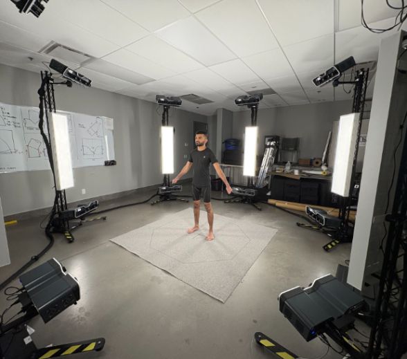
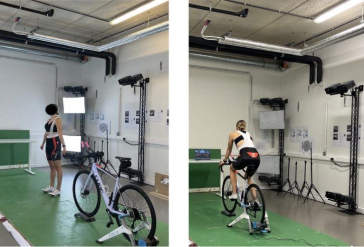
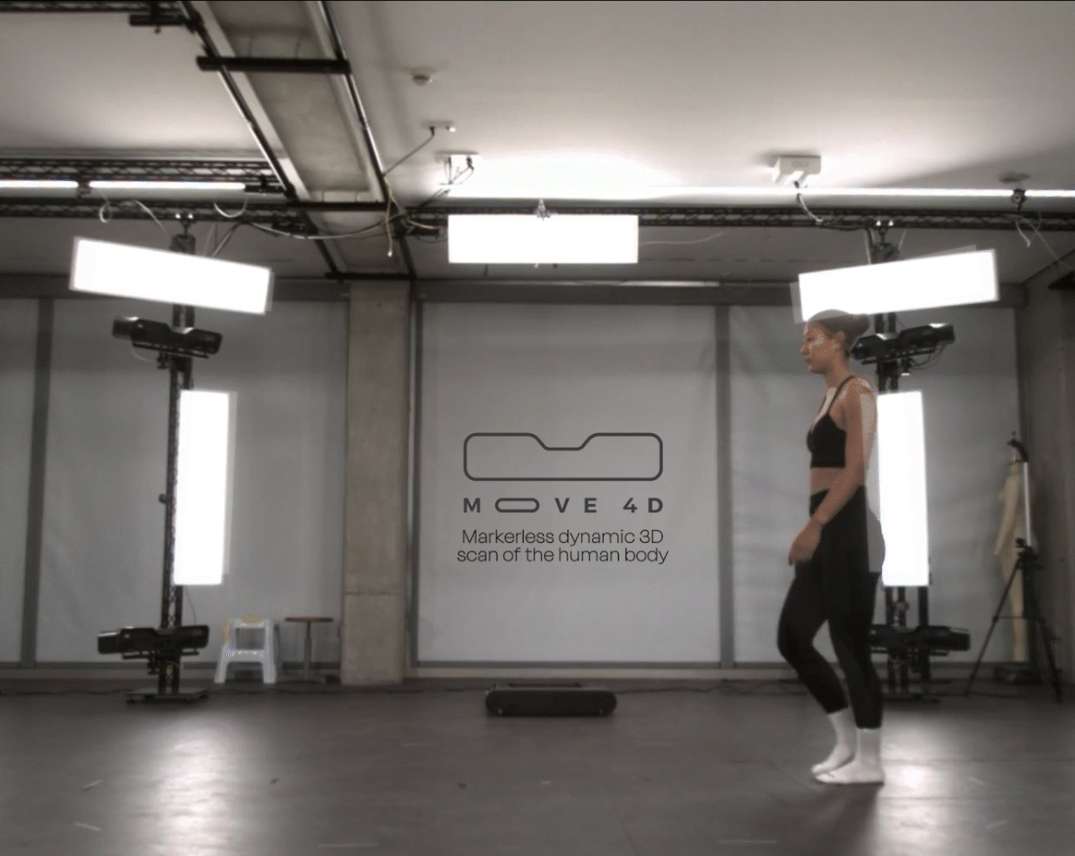
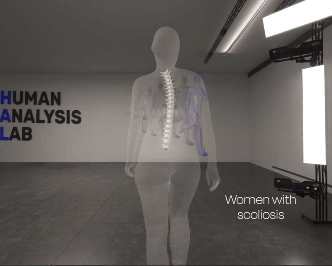
In this context, technologies such as MOVE4D, the high-precision markerless 4D body scanning system developed by the Instituto de Biomecánica (IBV), make it possible to analyze spinal morphology and movement simultaneously during functional tasks such as gait.
By capturing the external body surface in motion, 4D scanning allows clinicians and researchers to observe how spinal deformities influence functional activities like walking—without the need for markers or radiation.
From back surface to functional metrics: What 4D scanning reveals
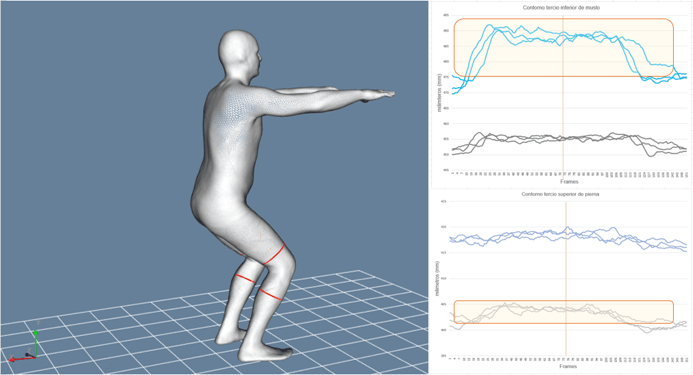
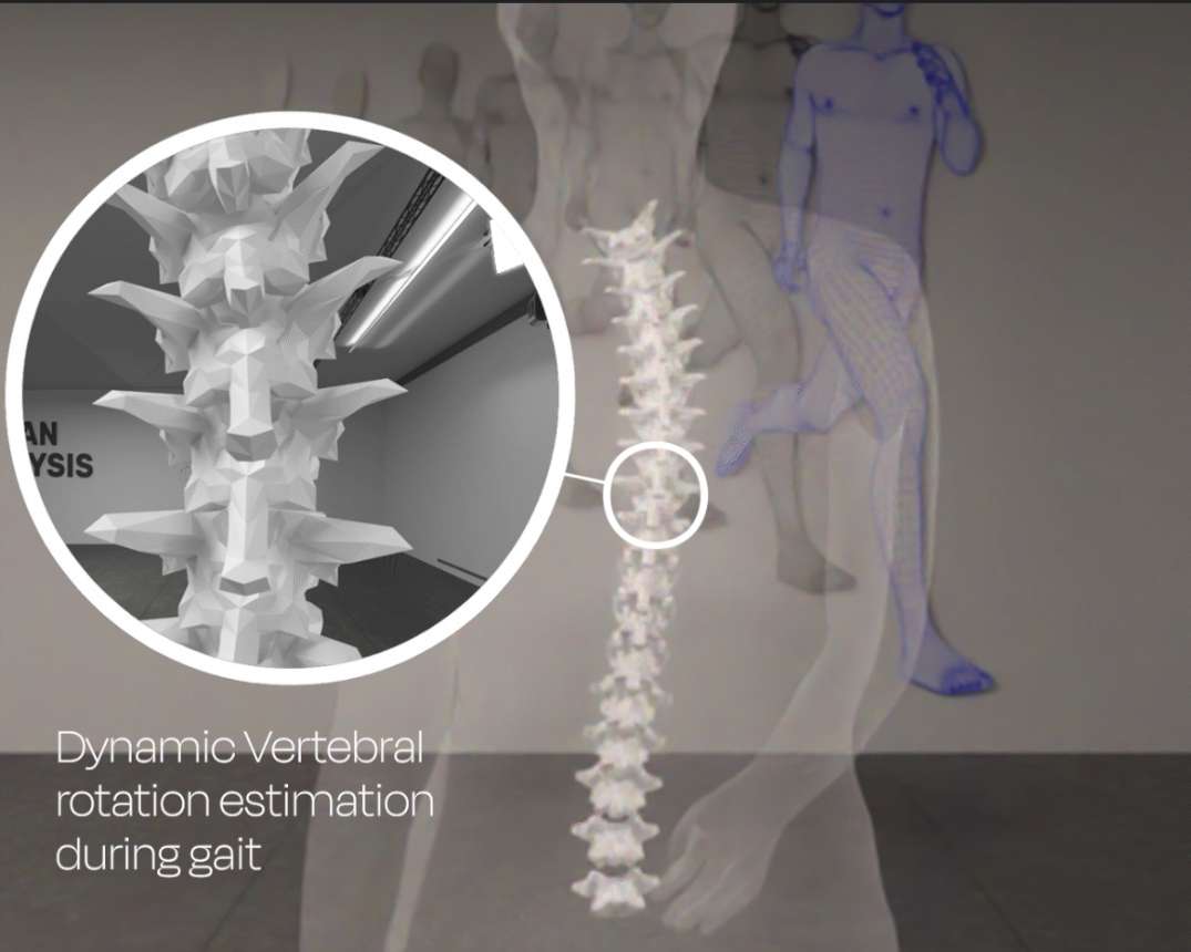
4D body scanning builds on traditional back surface analysis, extending it into the time domain. Instead of evaluating posture at a single static moment, this technology reconstructs the back surface continuously during gait, allowing estimation of the spinal midline and vertebral orientations from C7 to L5.
Through automated topological analysis of the back surface, 4D scanning extracts objective functional metrics that reflect how spinal deformity manifests during movement.
These metrics provide insight into compensatory strategies, asymmetries, and adaptations that cannot be identified through static assessment alone, making scoliosis assessment more comprehensive and clinically meaningful.
What advantages does 4D surface scanning offer over traditional methods?
Comparing 4D scanning and radiography for scoliosis monitoring
- Radiography remains the gold standard for measuring spinal curvature, but it presents two important limitations: exposure to ionizing radiation and a purely static representation of the spine. For long-term monitoring—especially in young patients—reducing radiation exposure is a critical concern.
- 4D surface scanning does not replace radiography but complements it. By offering a radiation-free, functional assessment, it allows clinicians to evaluate how spinal deformity affects movement, posture, and balance between radiographic follow-ups.
This combined approach enhances scoliosis assessment by linking structural information with real-world functional behavior.
Advantages over marker-based motion capture systems
- No manual marker placement and less operator dependency: Conventional motion capture systems used in instrumented gait analysis typically rely on markers placed on anatomical landmarks.
In patients with scoliosis, landmark identification can be challenging due to asymmetry, soft tissue artifacts, and operator dependency, which may reduce reliability and repeatability.
- Full-body surface capture with high spatial and temporal resolution: The technology captures the entire body surface with high spatial and temporal resolution, enabling automated and reproducible measurements without manual landmark placement.
This improves usability, reduces setup time, and increases consistency across sessions and operators.
Reducing patient exposure and improving comfort
Beyond accuracy, patient experience is a key advantage of 4D scanning. The non-invasive, markerless, and radiation-free nature of the system improves comfort and safety, making it particularly suitable for repeated assessments.
This is especially relevant for pediatric and adolescent scoliosis patients who require long-term follow-up.
Other articles that may interest you: MOVE4D at ESMAC 2025: Advancing the functional assessment of scoliosos
Which functional metrics can 4D scanning provide during gait?
1. Measuring spinal rotation, flexion, and lateral deviations
Using high-frequency dynamic captures during walking, 4D scanning enables the estimation of vertebral orientations and spinal motion in both frontal and transverse planes.
Metrics such as lateral flexion, axial rotation, and intervertebral motion patterns can be derived indirectly from back surface topology.
In the referenced study, differences in vertebral rotations differentiated the scoliosis case from healthy controls, highlighting the sensitivity of 4D scanning to subtle functional alterations associated with spinal deformity.
2. Tracking segmental movements in real-time
One of the strengths of 4D scanning is its ability to track segmental motion continuously. Variables such as trunk imbalance (TI) and acromion–pelvis angle (APA) were calculated throughout the gait cycle using homologous anatomical points on the mesh.
Results showed asymmetries in these gait-related parameters in the scoliosis case, with APA displaying asymmetric distributions not present in controls.
These findings demonstrate how 4D scanning captures dynamic compensations that emerge during locomotion.
3. Complementing instrumented gait analysis for full-body assessment
By integrating markerless 4D surface data from systems such as MOVE4D with instrumented gait analysis, clinicians can achieve a more comprehensive functional assessment of scoliosis.
Find all the applications for our product!
Rather than replacing instrumented gait analysis, 4D body scanning complements it.
While force plates and traditional kinematic systems quantify external loads and joint motion, 4D scanning adds detailed information on spinal deformity behavior and back surface dynamics.
This combined approach enables a more holistic functional assessment, linking spinal kinematics with gait parameters to better understand how scoliosis affects whole-body movement.
How can 4D scanning transform clinical and research applications in scoliosis?
Personalized functional assessment for patient-specific treatment
The ability to quantify functional adaptations and compensations opens new possibilities for personalized scoliosis management.
Clinicians can use 4D-derived metrics to monitor functional changes over time, evaluate treatment outcomes, and tailor interventions based on individual movement patterns rather than static images alone.
This functional perspective supports more informed decision-making in conservative treatment, rehabilitation, and follow-up strategies.
Future perspectives: From clinical evaluation to ergonomic design
Beyond clinical practice, 4D external body scanning has significant potential in research and applied fields.
By providing accurate and non-invasive, automated, and repeatable measurements, it enables large-scale studies on spinal deformity, movement variability, and adaptation mechanisms.
Future applications may extend to ergonomic design, assistive devices, and product development, where understanding how spinal deformities interact with movement is essential.
As markerless technologies continue to evolve, 4D scanning is poised to become a key tool in advancing both clinical scoliosis assessment and biomechanical research.
Want to learn more about MOVE4D and its clinical and research applications?
Visit MOVE4D or contact our team to discover how this technology is redefining motion analysis.